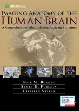
Imaging Anatomy of the Human Brain: A Comprehensive Atlas Including Adjacent Structures (en Inglés)
Reseña del libro "Imaging Anatomy of the Human Brain: A Comprehensive Atlas Including Adjacent Structures (en Inglés)"
"[A] very fine and detailed anatomic atlas... Dr. Borden et al. present an excellent atlas done basically by neuroradiologists…but with a very rich content, which is highly instructive also for neurologists and neurosurgeons, given the major importance of neuroradiological imaging in both practices."--Guilherme Carvalhal Ribas, MD, University of São Paulo Medical School, Neurosurgery"[A]n outstanding anatomic reference textbook... do yourself a favor and either purchase this book for your own library or select it for your department’s library. For proof of its value, examine it at the next ASNR or RSNA. You will be convinced. The bulk of the information in the book will never be out of date."--American Journal of Neuroradiology"I was very happy to be able to review this wonderful resource, which is packed with information and illustrations... The illustrations in this atlas are remarkable...I believe this text would be a valuable reference for any neurodiagnostic laboratory as neurodiagnostic techs move into more collaborative roles as part of the patient care team. I would highly recommend it." --Kathy Johnson, R. EEG/EP T., RPSGT, FASET, The Neurodiagnostic Journal"This atlas provides high quality images of the brain and adjacent structures in MR, CT, and neonatal ultrasound. The images are clearly labeled with a thorough index to allow for cross referencing between modalities... the inclusion of higher-end imaging such as fMRI, diffusion tensor imaging (DTI), and CT perfusion makes for a complete neuroanatomy atlas that will be useful for a long time... Weighted Numerical Score: 99 - 5 Stars!" -- Joel M Fritz, MD, Baystate Medical Center, Doody's Reviews"This volume presents a detailed and beautifully illustrated tour of neuroanatomy, employing…standard and advanced modalities… Ample use of color illustrates fiber tracts and differentiates different nuclei and other structures. The sections are amply labeled and easy to follow, allowing the reader to become familiar with cognate areas on images of multiple techniques." -- Carl E. Stafstrom, Division of Pediatric Neurology, Johns Hopkins Hospital, Journal of Pediatric Epilepsy An Atlas for the 21st Century The most precise, cutting-edge images of normal cerebral anatomy available today are the centerpiece of this spectacular atlas for clinicians, trainees, and students in the neurologically-based medical and non-medical specialties. Truly an "atlas for the 21st century," this comprehensive visual reference presents a detailed overview of cerebral anatomy acquired through the use of multiple imaging modalities including advanced techniques that allow visualization of structures not possible with conventional MRI or CT. Beautiful color illustrations using 3-D modeling techniques based upon 3D MR volume data sets further enhances understanding of cerebral anatomy and spatial relationships. The anatomy in these color illustrations mirror the black and white anatomic MR images presented in this atlas. Written by two neuroradiologists and an anatomist who are also prominent educators, along with more than a dozen contributors, the atlas begins with a brief introduction to the development, organization, and function of the human brain. What follows is more than 1,000 meticulously presented and labelled images acquired with the full complement of standard and advanced modalities currently used to visualize the human brain and adjacent structures, including MRI, CT, diffusion tensor imaging (DTI) with tractography, functional MRI, CTA, CTV, MRA, MRV, conventional 2-D catheter angiography, 3-D rotational catheter angiography, MR spectroscopy, and ultrasound of the neonatal brain. The vast array of data that these modes of imaging provide offers a wider window into the brain and allows the reader a unique way to integrate the complex anatomy presented. Ultimately the improved understanding you can acquire using this atlas can enhance clinical understanding and have a positive impact on patient care. Additionally, various anatomic structures can be viewed from modality to modality and from multiple planes. This state-of-the-art atlas provides a single source reference, which allows the interested reader ease of use, cross-referencing, and the ability to visualize high-resolution images with detailed labeling. It will serve as an authoritative learning tool in the classroom, and as an invaluable practical resource at the workstation or in the office or clinic. Key Features: Provides detailed views of anatomic structures within and around the human brain utilizing over 1,000 high quality images across a broad range of imaging modalities Contains extensively labeled images of all regions of the brain and adjacent areas that can be compared and contrasted across modalities Includes specially created color illustrations using computer 3-D modeling techniques to aid in identifying structures and understanding relationships Goes beyond a typical brain atlas with detailed imaging of skull base, calvaria, facial skeleton, temporal bones, paranasal sinuses, and orbits Serves as an authoritative learning tool for students and trainees and practical reference for clinicians in multiple specialties

